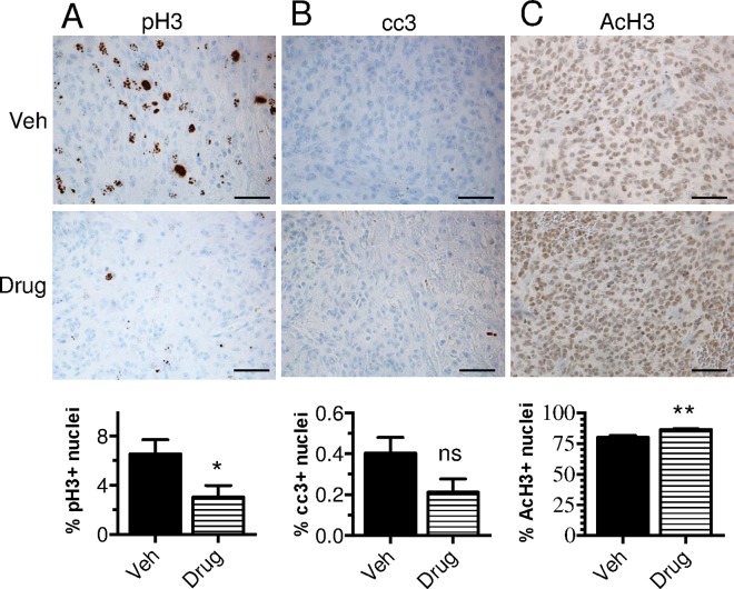
Fig 5. Short-term in vivo treatments with panobinostat reduces tumor cell proliferation and increases H3 acetylation without evidence of apoptosis.
Neonatal Ntv-a;p53-fl/fl mice were injected with RCAS-PDGF-B, -H3.3-K27M, and–Cre viruses to induce tumor formation. Upon the first appearance of brain tumor symptoms (3–5 weeks post-injection), mice were treated with five doses of 20 mg/kg panobinostat (Drug) or vehicle (Veh, 25% DMSO, 0.25x PBS, 5% glucose, n = 6 in each group) once daily via i.p. injections and then sacrificed 1 hour after the final treatment. Shown is immunohistochemistry of brain tumor tissue for cell proliferation (phospho histone H3, pH3) (A), cell apoptosis (cleaved caspase-3, cc3) (B), and H3 acetylation (AcH3) (C), 400x, scale bar = 50 μm, with quantification of total positive nuclear staining as a percentage of total nuclear area below in each panel. Statistical significance was determined using unpaired two-tailed t-test to compare groups. (ns: nonsignificant).

