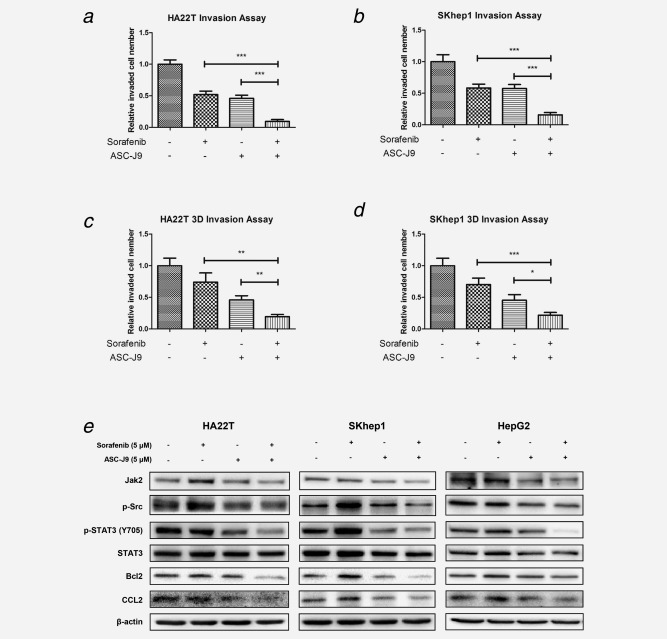
Figure 3.

ASC‐J9® enhanced Sorafenib efficacy to suppress HCC cell invasion. (a, b) Chamber‐transwell invasion assays were performed. 3 × 10⁴ HA22T cells or 5 × 10⁴ SKhep1 cells in serum free DMEM were plated into the upper chambers and 600 μl 10% FCS medium was placed in the lower chambers for incubation at 37°C in 5% (v/v) CO2 incubator for 24 hr. The invaded cells (to lower membrane surface) were counted in five randomly chosen microscopic fields (100×) of each experiment and pooled for statistical analysis. (c, d), 3D‐invasion assays were performed using HA22T and SKhep1 cells. The cells with protrusions were regarded as invaded cells and 10 random different fields under 200× magnification were counted for quantification. Each sample was run in triplicate and in multiple experiments. p < 0.05 was considered statistically significant. *p < 0.05, **p < 0.01 and ***p < 0.005. (e), ASC‐J9® combined with Sorafenib synergistically suppressed HCC cell invasion and proliferation via altering p‐STAT3/CCLs and p‐STAT3/Bcl‐2 signals in three different HCC cell lines. HCC cells were plated in 6‐well plates at 2 × 10⁵ (HA22T) or 5 × 10⁵ (SKhep1 and HepG2) cells/well and treated as designated for 48 hr. After treatments, the cells were lysed and total protein was extracted for Western blot assay, which showed that Sorafenib and ASC‐J9® combination therapy significantly suppressed p‐STAT3/CCL2 signals compared with ASC‐J9® or Sorafenib alone in all three cell lines. Three different independent experiments were performed.
