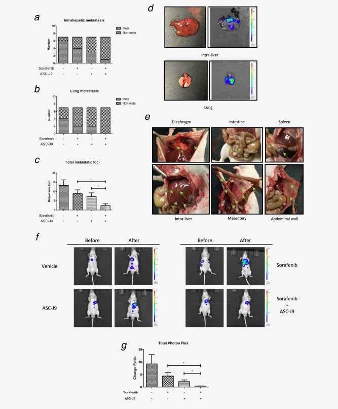
Figure 5.

ASC‐J9® combined with Sorafenib better suppressed HCC growth and metastasis in vivo. (a, b) IVIS imaging was used to determine the intrahepatic metastasis and lung metastasis. (c) Total metastatic foci were counted by autopsy. (d) Representative bioluminescent images of intrahepatic metastasis and lung metastasis. (e) Representative images of diaphragm, intestine, spleen, intra‐liver, mesentery and abdominal wall metastatic foci (green arrows). (f) Representative bioluminescent images before and after treatment in different treatment groups. (g) The change folds of total photon flux after treatment comparing to total photon flux before treatment were calculated using Living Image® software (PerkinElmer).
