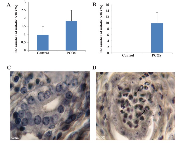
Fig.3.
Up-Statistical comparison between control and estradiol valerate (EV)-treated polycystic ovary syndrome (PCOS) rats. A. The number of mitotic cells in the luminal epithelium (P<0.05), B. The number of mitotic cells in the glandular epithelium (P<0.001). Down-Histological sections of the uterus from control and EV-treated PCOS rats following iron hematoxylin (iron-H) staining, C, and D. The glandular epithelium (×1000). ↗; Mitotic cells

