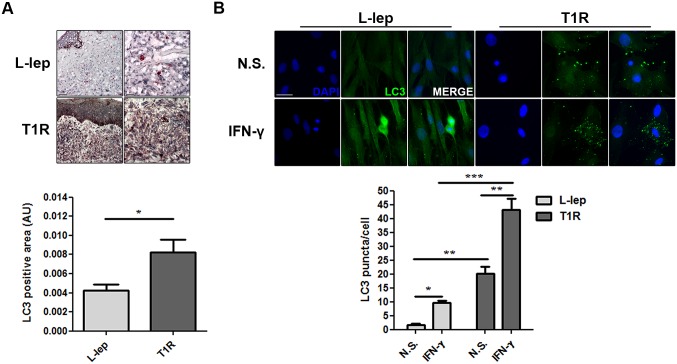
Fig 8. T1R episodes enhances the levels of autophagy in L-lep patients.
(A) Immunohistochemistry (IHC) for endogenous LC3. Increase of the LC3 expression in skin lesion cells of L-lep patients undergoing T1R episodes. IHC images were quantified and data are expressed as arbitrary units (AU). Bars represent the mean values ± SEM (L-lep, n = 4; T1R, n = 3). *P < 0.05, Mann-Whitney test. Scale bars: 50 and 25 μm. (B) Macrophages (MΦs) were isolated from skin lesions of L-lep and T1R patients and treated with IFN-γ (10 ng/mL) for 18 h. Cells were fixed and stained with the anti-LC3 antibody (green) and DAPI (blue). Non-stimulated (N.S.) T1R MΦs showed more enhanced LC3 puncta formation than L-lep MΦs. IFN-γ stimulation led to accumulation of LC3 dots in both L-lep and T1R MΦs, but to a lesser extent in MΦs derived from L-lep patients without T1R lesions. Immunofluorescence images were quantified and bars represent the mean values of the number of LC3 puncta per cell ± SEM of three independent experiments. *P < 0.05, **P < 0.01, **P < 0.001, Mann-Whitney test. Scale bar: 50 μm.

