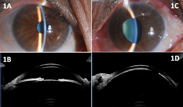
Fig 1. Anterior segment photograph and UBM images of a 46-year-old female patient.
(1A, 1B) Photograph and UBM showed relatively shallow anterior chamber of the right eye. (1C) Color photograph showed flat anterior chamber and moderately dilated pupil of the left eye. (1D) UBM findings showed flat anterior chamber (nearly corneo-lenticular touch) in the left eye one month after trabeculectomy.

