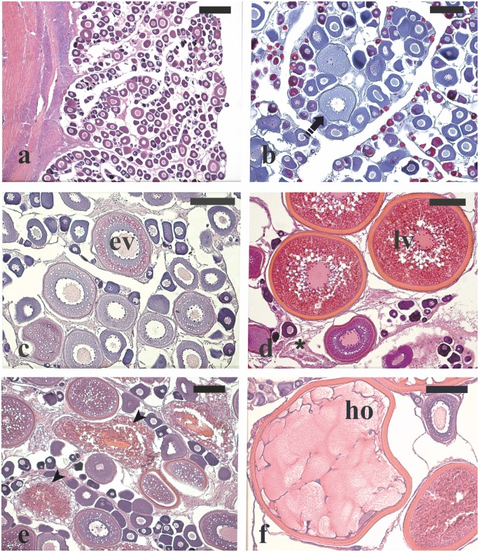
Fig 2. Micrographs of ovary sections from female greater amberjack sampled in three different phases of the reproductive season.
(a) Wild individual sampled on 1 May showing perinucleolar oocytes as the most advanced stage in the ovary. (b) Cortical alveoli oocytes in the ovary of a wild specimen captured on 1 May 2015. (c) Early vitellogenic oocytes in the ovary of a wild individual sampled on 1 May 2015. (d) Late vitellogenic oocytes together with post-ovulatory follicles from a wild spawning fish caught on 31 May 2014. (e) Extensive atresia of late vitellogenenic follicles in a captive-reared specimen sampled on 4 June 2015. (f) Hydrated oocyte from a spawning wild fish sampled on 30 June 2014. Haematoxylin-eosin staining in (a), (c), (d), (e) and Mallory’s trichrome staining in (b). Magnification bars = 300 μm in (a) and 150 μm in (b)-(f). Arrowhead: atretic late vitellogenic follicle; asterisk: post-ovulatory follicle; dashed arrow: cortical alveoli stage oocyte; ev: oocyte in early vitellogenesis stage; ho: hydrated oocyte; lv: oocyte in late vitellogenesis stage.

