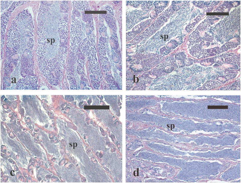
Fig 5. Micrographs of testes sections from male greater amberjack sampled in three different phases of the reproductive season.
(a) Testis section from a wild individual sampled on 1 May showing the presence of all stages of spermatogenesis in the germinal epithelium and a limited amount of luminal spermatozoa. (b) Testis section from a wild fish caught on 31 May 2014, showing all stages of spermatogenesis as well as large amount of luminal spermatozoa. (c) Testis section from a captive-reared fish sampled on 4 June 2015 showing an arrested spermatogenesis state, with residual sperm cysts in the germinal epithelium and abundant spermatozoa in the lumen of seminiferous lobules. (d) Testis sections from a captive-reared specimen caught on 2 July 2015 showing a moderate amount of spermatozoa in the lumen of seminiferous lobules. Haematoxylin-eosin staining. Magnification bars = 100 μm in (a) and (b), 200 μm in (c) and (d). sp: spermatozoa in the lumina of seminiferous lobules.

