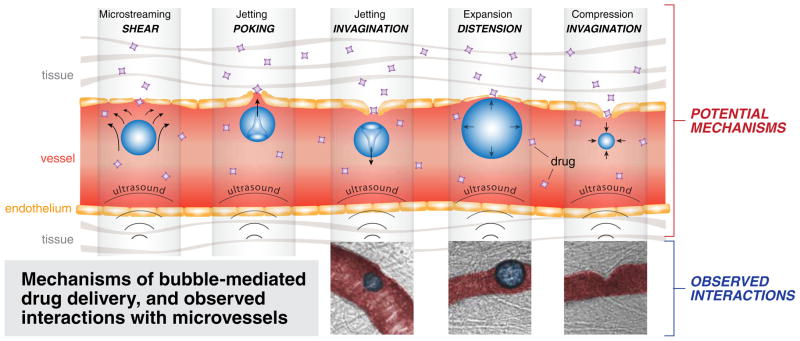
Fig. 1.
Schematic and ex vivo microscopy images of non-thermal vascular effects of therapeutic ultrasound. Top row demonstrates microstreaming (a), jetting (b, c), bubble expansion/compression (d, e). Bottom row images demonstrate interaction of lipid-coated perfluoropropane ultrasound contrast bubbles with capillary walls (injected with Green India Ink for contrast) in ex vivo rat mesentery, during HIFU treatment, under 40× high-speed microscopy [9, 10]

