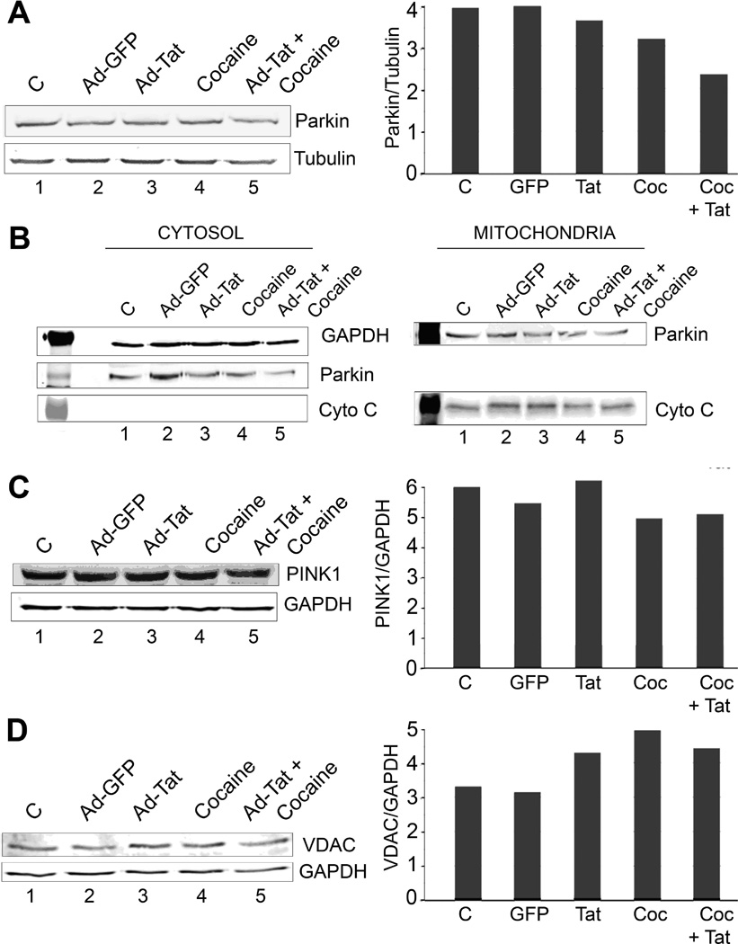
Figure 3. Expression of VDAC, PINK1 and Parkin in primary hippocampal neurons treated with HIV-1 Tat and cocaine, alone or in combination.
Rat primary hippocampal neurons were harvested from E18 rat embryos and plated on Poly-D-Lysine-coated 12-well plates. Cells were then transiently transduced for 2 h with either Adeno-GFP or Adeno-Tat (moi = 1). After 48h, the cultured neurons were treated with 1 µM cocaine in sodium citrate (pH 5.0) or 10 µM ionomycin as a positive control. Western blots were performed for Parkin (A). To examine the subcellular distribution of Parkin, the cultured neurons were fractionated into cytosol and mitochondrial fractions as described and analyzed by Western blot for Parkin, cytochrome c (mitochondrial marker) and GAPDH (cytosolic marker) as shown in Panel B. Western blots of total cell extracts were also performed for PINK1 (C) and VDAC (D). Quantification for each Western is shown in the right hand side of Panels A, C and D. Parkin is slightly downregulated in cocaine-treated neurons and there is translocated from cytosol to mitochondria after treatment with Tat and cocaine. PINK1 is also slightly downregulated with cocaine alone and cocaine plus Tat. VDAC was upregulated by both Tat and cocaine suggesting a possible response to neuronal stress and may indicate increased mitophagy signaling.

