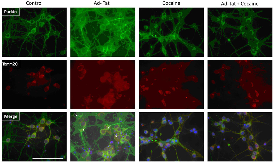
Figure 4. Analysis of the effect of HIV-1 Tat and cocaine, alone or in combination on Parkin and Tomm20 subcellular distribution in primary hippocampal neurons by immunocytochemistry.
Primary rat hippocampal neurons were harvested from E18 rat embryos and plated on Poly-D-Lysine-coated chamber slides. Cells were then transiently transduced for 2 h with either Adeno-GFP or Adeno-Tat (moi = 1). After 48h, the cultured neurons were treated with 1 µM cocaine in sodium citrate (pH 5.0). For labeling of Parkin (green) and Tomm20 (a mitochondrial marker, red), immunocytochemistry was performed as described in Materials and Methods with rabbit anti-Tomm20 (1:1000) and mouse anti-Parkin (1:1000). Nuclei are stained with DAPI (blue). Arrows indicate endogenous Parkin-ring-like structures surrounding fragmented mitochondria. Parkin-ring-like structures were visible around fragmented mitochondria of cells treated with Tat. The scale bar is 40 μm.

