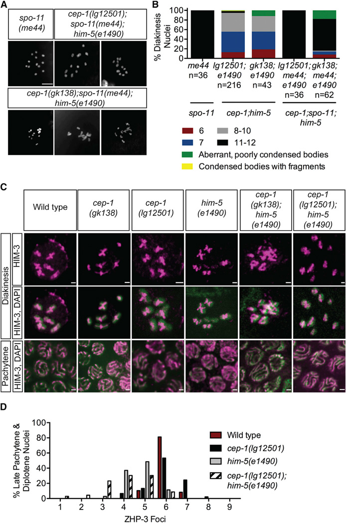
Figure 3. cep-1 and him-5 Promote Crossover Formation.
(A) Representative confocal images of DAPI-stained diakinesis chromosomes of young adult animals of the indicated genotypes. The scale bar represents 5 µm. See also Figures S3A and S3B.
(B) Quantification of DAPI-stained bodies in diakinesis oocytes of young adult animals of the indicated genotypes. n, number of oocytes. Data for cep-1;him-5 double mutants were taken from Figure 1D.
(C) Pachytene and diakinesis nuclei of wild-type (1), cep-1(gk138) (2), cep-1(lg12501) (3), him-5(e1490) (4), cep-1(gk138);him-5(e1490) (5), and cep-1(lg12501);him-5(e1490) (6) stained with antibody against the chromosome axis protein HIM-3. See also Figures S3C–S3G.
(D) Frequency distribution of ZHP-3 foci in late pachytene and diplotene nuclei of the indicated genotypes. Statistical comparisons between genotypes were performed using the two-tailed Mann-Whitney test; 95% CI. Wild-type versus him-5(e1490), p < 0.0001;him-5(e1490) versus cep-1(lg12501);him-5(e1490), p = 0.0070.

