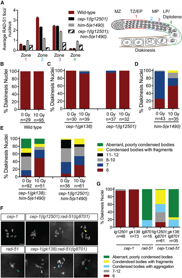
Figure 4. cep-1 and him-5 Regulate Meiotic DSB Formation.
(A) Quantification of RAD-51 foci in the meiotic germlines of worms of the indicated genotypes and schematic of the C. elegans germline. Each gonad was divided into four meiotic zones (TZ/EP, transition zone/early pachytene; MP, mid-pachytene; LP/diplotene, late pachytene). Error bars represent SD. See also Table S2.
(B) Quantification of DAPI-stained bodies in diakinesis oocytes of wild-type worms 24 hr post-IR treatment. See also Figure S4A.
(C) Quantification of DAPI-stained bodies in diakinesis oocytes of cep-1(gk138) and cep-1(lg12501) single mutants 24 hr post-IR treatment. See also Figure S4B.
(D) Quantification of DAPI-stained bodies in diakinesis oocytes of him-5(e1490) single mutants 24 hr post-IR treatment. See also Figure S4C.
(E) Quantification of DAPI-stained bodies in diakinesis oocytes of cep-1(gk138);him-5(e1490) and cep-1(lg12501);him-5(e1490) double mutants 24 hr post-IR treatment. See also Figure S4D.
(F) Representative confocal images of DAPI-stained diakinesis chromosomes of adult animals of indicated genotypes. The scale bar represents 5 µm. Image for cep-1 single mutant was taken from Figure 1C. Green triangle indicates aberrant, poorly condensed chromosomes. Yellow triangle indicates chromosome fragments. Light blue triangle indicates condensed bodies with aggregations. See also Figure S1.
(G) Quantification of DAPI-stained bodies in diakinesis oocytes of young adult animals of indicated genotypes. n, number of oocytes. Data for cep-1 single mutants were taken from Figure 1C.

