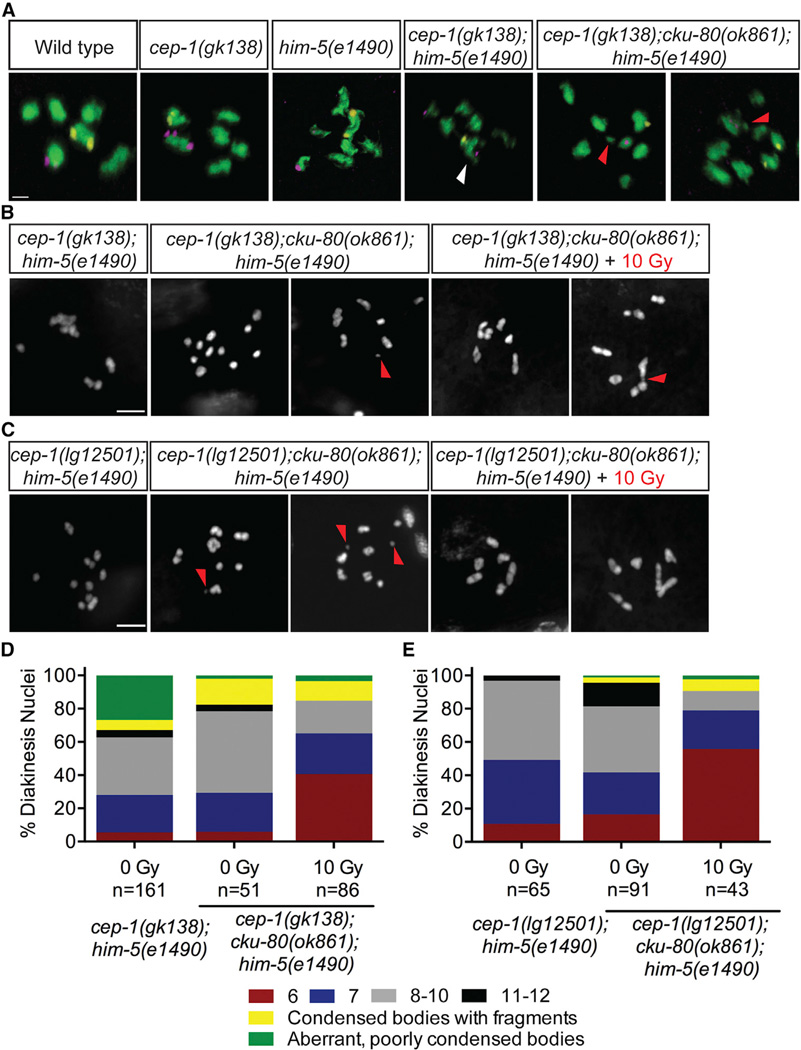
Figure 5. cep-1 and him-5 Prevent Inappropriate NHEJ Activity.
(A) Diakinesis nuclei of day 4 animals of the indicated genotypes labeled with FISH probes to chromosomes V (yellow) and X (magenta). DNA is shown in green. White arrow indicates a single DAPI body containing both FISH probes, demonstrating a fusion between nonhomologous chromosomes. Red triangle indicates chromosome fragments. White triangle indicates a single DAPI body containing probes for chromosomes V and X. The scale bar represents 1 µm. See also Table S3.
(B) Representative confocal images of DAPI-stained diakinesis chromosomes of cep-1(gk138);cku-80(ok1861);him-5(e1490) triple mutants 24 hr after mock (0 Gy) or IR (10 Gy) treatment. Image of cep-1(gk138);him-5(e1490) mutant is shown for comparison. Scale bar represents 5 µm. See also Figures S5A–S5C.
(C) Representative confocal images of DAPI-stained diakinesis chromosomes of cep-1(lg12501);cku-80(ok1861);him-5(e1490) triple mutants 24 hr after mock (0 Gy) or IR (10 Gy) treatment. Image of cep-1(lg12501);him-5(e1490) mutant is shown for comparison. Scale bar represents 5 µm. See also Figures S5A–S5C.
(D) Quantification of DAPI-stained bodies in diakinesis oocytes of cep-1(gk138);him-5(e1490) double and cep-1(gk138);cku-80(ok861);him-5(e1490) triple mutants 24 hr after mock (0 Gy) or IR (10 Gy) treatment.
(E) Quantification of DAPI-stained bodies in diakinesis oocytes of cep-1(lg12501);him-5(e1490) double and cep-1(lg12501);cku-80(ok861);him-5(e1490) triple mutants 24 hr after mock (0 Gy) or IR (10 Gy) treatment.

