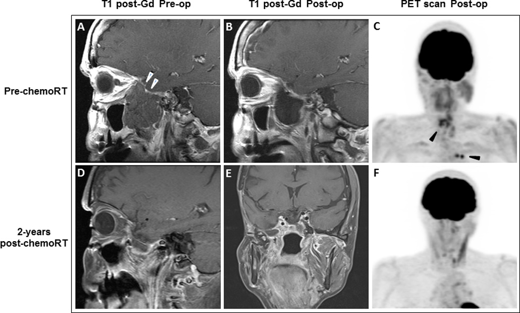
Figure 1. Case 1: Radiologic findings of surgical site and extraneural metastases before and after chemoradiation with temozolomide.

(A) Pre-operative sagittal image with T1-weighted gadolinium-enhanced MRI demonstrates a non-enhancing mass measuring 4.7 x 3.7 x 5.4 cm (white arrowheads) involving the lateral wall of the right sphenoid sinus, nasopharynx, right middle cranial fossa, and masticator space. (B) MRI shows postoperative changes in the right masticator space. All of the previously seen masticator space disease has been resected. A large fluid space is present below the temporal lobe. (C) PET scan for treatment planning purposes shows FDG-avid soft tissue density in the left posterior ethmoid air cells and the region of the right sphenoid sinus (suspicious for residual carcinoma) as well as in the mediastinum and hilar lymph nodes (black arrowheads). MRI (D and E) and PET scan (F) 2 years after chemoradiation shows stable findings in the masticator space and reduction of activity in the left posterior ethmoid sinus as well as the mediastinal and left hilar nodes.
