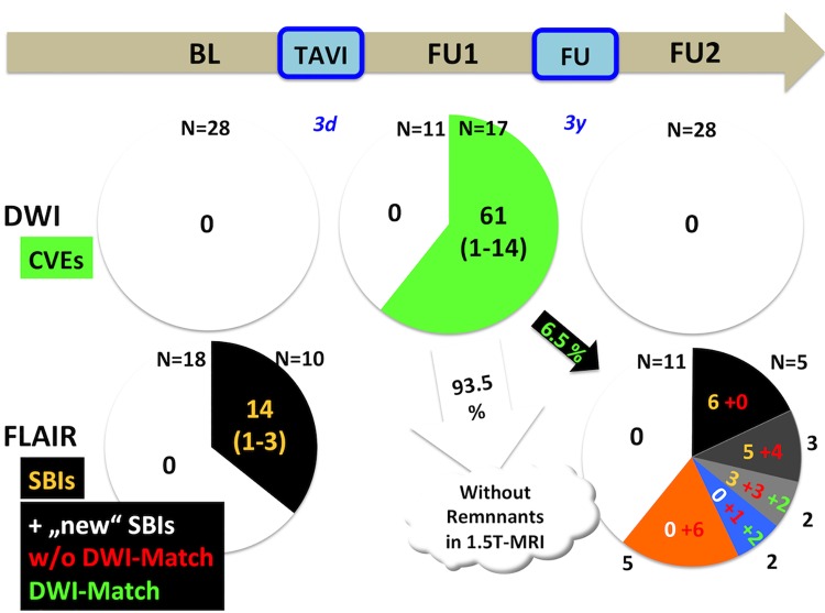
Fig 2. Trajectories of acute and subacute cerebral lesions after TAVI.
Patients underwent MRI prior (left), early (mid) and late (right) after TAVI. Above: The majority of patients demonstrate cerebral embolic events early after TAVI. No spontaneous lesions were observed prior and late after TAVI. Below: Ten out of 28 patients undergoing TAVI had 14 (old, non-procedural) silent brain infarctions (SBIs) in MRI (left). After long-term follow-up, seven patients without "old" SBIs revealed "new", post-procedural events. Notably, only four out of 18 "new" SBIs had an early procedural correlate in diffusion-weighted imaging (right).

