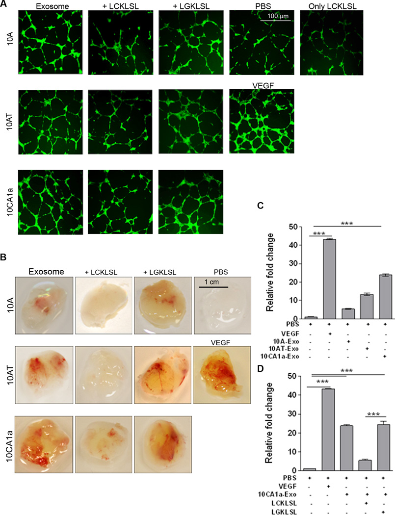
Figure 2. Exo-AnxA2 promotes angiogenesis.
Effect of exo-AnxA2 on in vitro tube formation assay. A) Fluorescent images of in vitro endothelial tube formation assay with HUVEC cells; (5 different fields per group were considered. Repeated in triplicates). PBS (negative control), VEGF (100ng/ml, positive control) and 100 µg of exosomal proteins were used (n=3). Peptide concentration used: 5µM (n=3). Exosomes from MCF10A, MCF10AT and MCF10CA1a cells are designated as 10A, 10AT and 10CA1a respectively. Scale bar 100 µm.
Matrigel Plug assay: Mice were injected with matrigel in the presence of PBS alone (negative control), VEGF alone (100 ng/ml, positive control), or 100 µg exosomes ± peptide. After 12 days, the plugs were removed and the angiogenic responses were evaluated. D) Representative images of matrigel plugs are shown. Scale bar 1 cm. Hemoglobin estimation of homogenized matrigel plugs by Drabkin’s method (C–D). Fold change to PBS is shown. Peptide concentration: 5µM (n=3). Exosomes from MCF10A, MCF10AT, and MCF10CA1a cells are designated as 10A, 10AT, and 10CA1a, respectively. (*) p < 0.05, (**) p< 0.01, (***) p < 0.001, (****) p < 0.0001.

