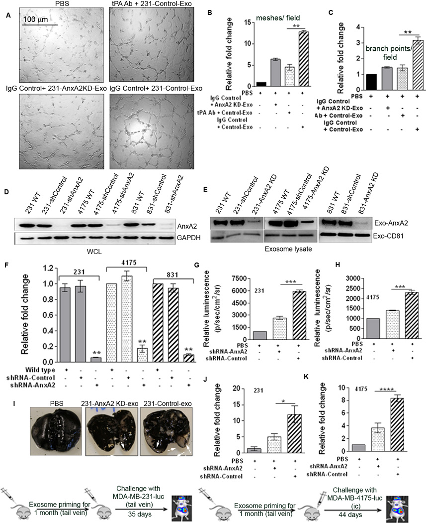
Figure 3. Exo-AnxA2 promotes angiogenesis via tPA and activates macrophages, leading to secretion of IL-6 and TNF-alpha.
(A) Endothelial tube formation assay showing the role of tPA in the pro-angiogenic effect of exo-AnxA2 (n=2). Quantification of the number of meshes/field (B) and number of branch points/field (C).
Exo-AnxA2 promotes breast cancer metastasis to lungs . Western blot analysis of WCL (D) and exosomal lysates (E) showing knockdown of AnxA2 (n=2); GAPDH and CD81 were used as loading controls for (D) and (E), respectively. Quantification of exosomal AnxA2 (F). G) Quantification of BLI of PBS-, 231-AnxA2KD-Exo-, or 231-Control-Exo-primed animals 35 days after challenge with MDA-MB-231-luc cells (lateral tail vein injection), showing differences in the extent of lung metastasis (n=8). Fold change in photon flux to PBS-primed animals is shown. H) Quantification of bioluminescence (BLI) of PBS-, 4175-AnxA2KD-Exo-, or 4175-Control-Exo-primed animals 44 days after challenge with MDA-MB-4175-luc cells (intracardiac; ic) showing differences in the extent of lung metastasis (n=8). Fold change in photon flux to PBS-primed animals is shown. I) India ink staining of the excised lungs from MDA-MB-231-luc cell-injected animals, showing the number of metastatic nodules. Quantification of the number of metastatic lung nodules with MDA-MB-231-luc (tail vein injection) treatment (J) and MDA-MB-4175-luc (ic) treatment (K). Fold change compared to PBS-primed animals is shown. (*) p < 0.05, (**) p< 0.01, (***) p < 0.001, (****) p < 0.0001.

