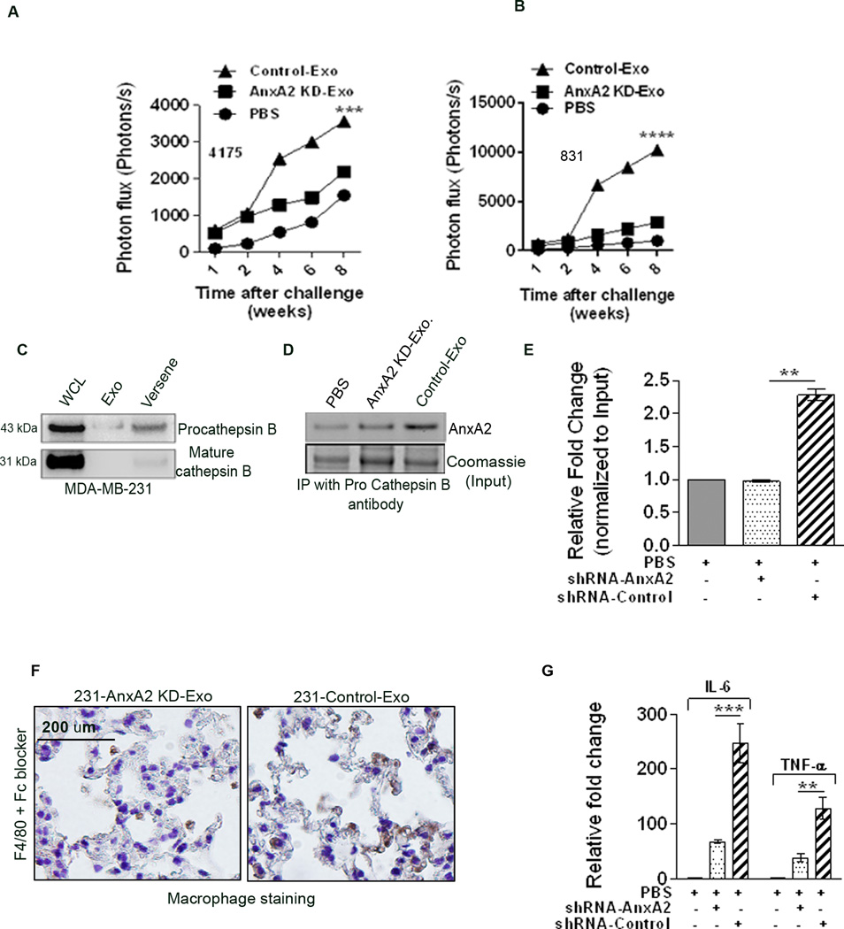
Figure 5.
Quantification of photon flux change over time: (A) MDA-MB-4175-luc-injected lung metastasis and (B) MDA-MB-831-luc-injected brain metastasis. C) Western blot analysis showing the expression of pro-cathepsinB and mature cathepsinB in WCL, Exo lysate, and membrane wash (Versene) fractions. D) Immunoprecipitation of AnxA2 with pro-cathepsinB antibody in the membrane wash lysate after treatment with PBS, AnxA2KD-Exo, or Control-Exo. E) Densitometric analysis of the blot (n=2). (*) p < 0.05, (**) p< 0.01, (***) p < 0.001, (****) p < 0.0001. DAB immunostaining of macrophages in the lung sections with F4/80 antibodies in 231-AnxA2KD-Exo- and 231-Control-Exo-primed animals (F) after Fc blocking. Scale bar = 100 µm. G) ELISA of lung extracts to analyze IL-6 and TNF-alpha levels from PBS-, 231-AnxA2KD-Exo-, and 231-Control-Exo-primed animals, 24 hrs after injection (n=2). (*) p < 0.05, (**) p< 0.01, (***) p < 0.001, (****) p < 0.0001.

