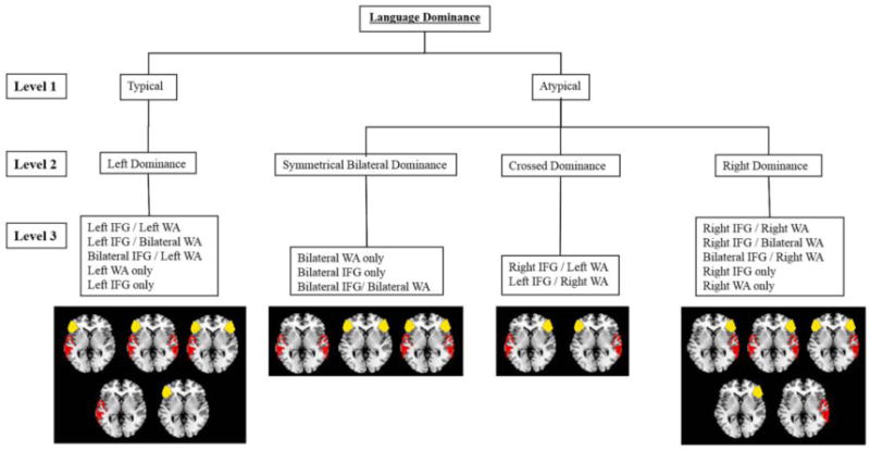
Figure 1.

Hierarchy of 15 typical and atypical language patterns based on categorical criteria in both patients and controls. Language is classified into 3 levels: typical=atypical, language dominance (left, bilateral, crossed, or right), and individual activation patterns. Regions of interest are left lateralized if lateralization index (LI) ≥ 0.20, bilateral if LI < |0.20|, and right lateralized if LI ≤ −20.20. IFG = inferior frontal gyrus; WA = Wernicke area. Figure modified with author's permission.
