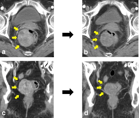
Fig. 1.

a Coronal CT images showed an enhanced tumor measuring 5.3 × 4.2 cm behind the rectum. b After the treatment of imatinib, coronal CT images revealed that the tumor size decreased to 3.5 cm. c Axial CT images showed an enhanced tumor at the right posterior side of the rectum. d After treatment of imatinib, axial CT images revealed that the tumor size decreased to 3.5 cm
