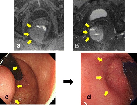
Fig. 2.

a T1-weighted MRI showed a low intensity tumor. b T2-weighted MRI showed a high intensity tumor. c Colonoscopic examination revealed a submucosal tumor at the posterior rectal wall, and that the distal tumor margin was 1 cm above the dentate line. d Colonoscopic examination revealed that the tumor markedly decreased after the treatment of imatinib
