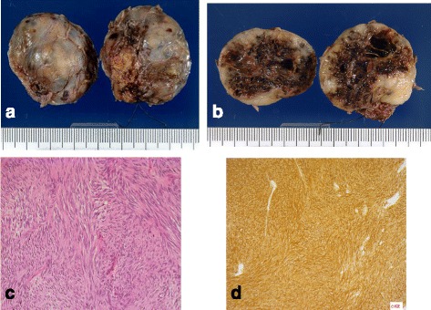
Fig. 4.

a, b The resected tumor was 3.5 × 3.5 × 2.5 cm in size. The rubbery-hard tumor with widespread central necrosis was completely capsulized. c Microscopic examination (hematoxylin-eosin staining, original magnification: ×400) revealed a formation of spindle-shaped cells. d Immunohistochemical staining of the tumor cells for c-kit revealed strong positive findings
