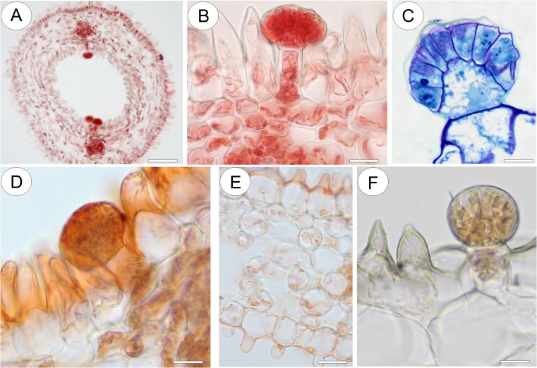
Fig. 12.
Anatomy and histochemistry of spur of Utricularia nelumbifolia. a Apical part of spur stained with Ponceau 2R. Glandular trichome distribution coincides with the position of vascular bundles; bar = 150 μm. b Head cells of glandular trichome stained intensely with Ponceau 2R; bar = 20 μm. c Detail of the head of glandular trichome following staining with MB/AII. Note the dense cytoplasm of the secretory head cells and that the cuticle has become detached from the outer cell walls; bar = 14 μm. d Cuticle overlying the glandular trichome stained uniformly with Sudan III; bar = 20 μm. e Epidermal and parenchyma cells of the spur contain small lipid droplets; bar = 30 μm. f Testing with IKI did not indicate the presence of starch in the conical cells of the epidermis nor the glandular trichomes; bar = 20 μm

