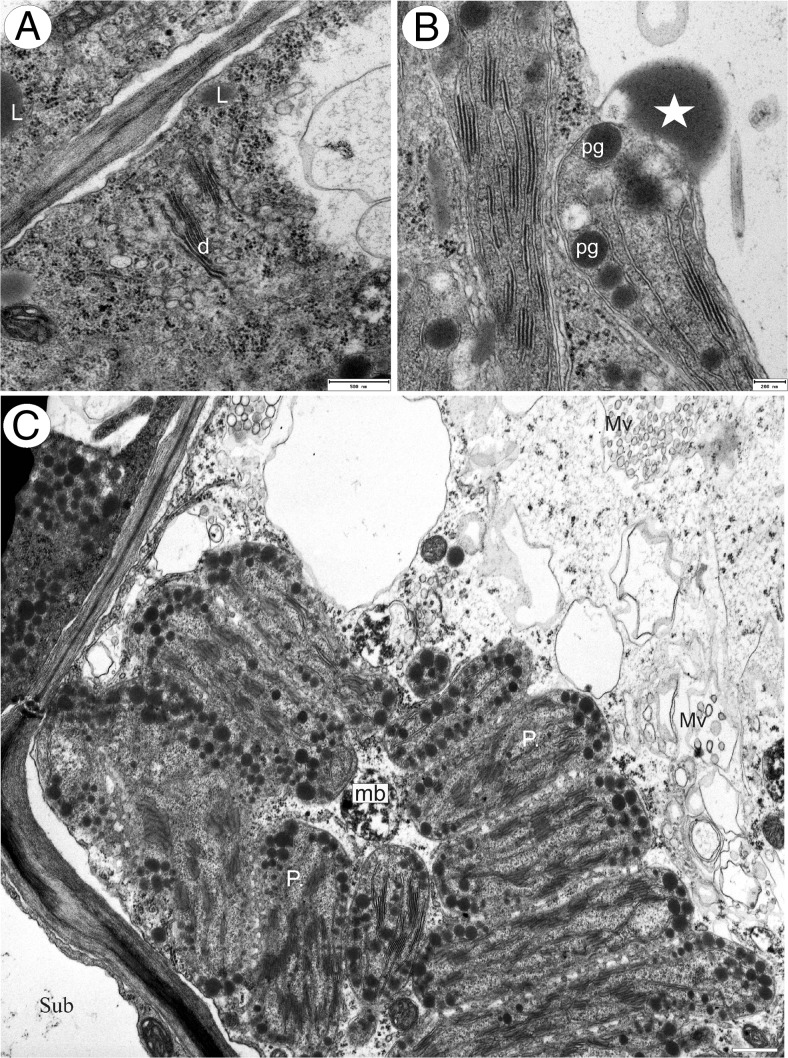
Fig. 7.
Ultrastructure of Utricularia cornigera palate papillae. a Electron micrograph showing dictyosomes (d) with numerous small vesicles and cytoplasmic lipid bodies (L); bar = 500 nm. b Plastids containing numerous, large plastoglobuli. Note the osmiophilic body (star) associated with plastid; bar = 200 nm. c General ultrastructure of the basal part of papilla. Note the multi-vesicular bodies (Mv), microbodies (mb), plastids (P), and part of subepidermal cell (Sub); bar = 0.9 μm

