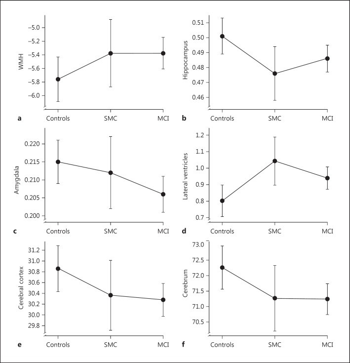
Fig. 1.
Mean age- and sex-adjusted ICV-corrected volumes of WMH (a), hippocampus (b), amygdala (c), lateral ventricles (d), cerebral cortex (e), and cerebrum (f) in the control, SMC and MCI groups. Lateral ventricles and WMH are log transformed. ICV-corrected volumes = (sum of bilateral volumes × 100) divided by total ICV. Error bars represent 95% confidence intervals.

