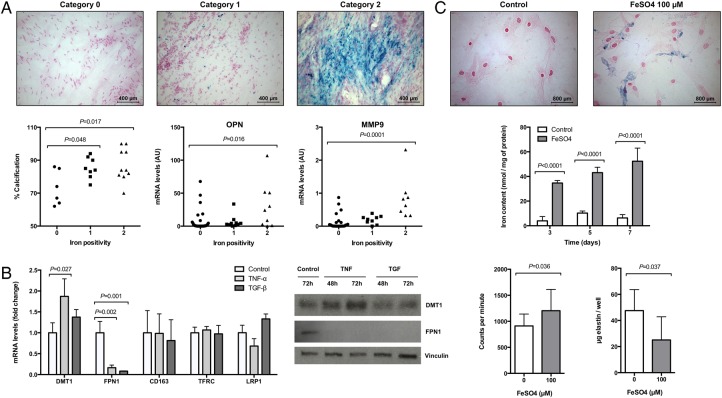
Figure 1.
Iron accumulation in stenotic valves is associated with calcification and affects valvular interstitial cells function. (A) Histological classification of iron positivity in human aortic valves using Perls' staining and its relation with calcification and gene expression. (B) Changes in mRNA and protein expression in valvular interstitial cells after stimulation with tumour necrosis factor-α and transforming growth factor-β for 24 or 48–72 h, respectively (n = 3). (C) Intracellular iron detection and quantification in valvular interstitial cells incubated with FeSO4 (100 µmol/L) for 72 h (n = 3) and its effect in 3H-thymidine incorporation (expressed in counts per minute; n = 3) and elastin production (n = 6). Multiple comparisons show adjusted P-values.

