Fig. 2.

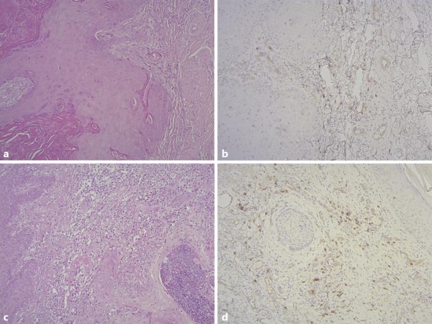
Histological pictures of keratoacanthoma. Haematoxylin and eosin stain (a) as well as CD123 staining (b) of a mature keratoacanthoma. Haematoxylin and eosin stain (c) as well as CD123 staining (d) of a keratoacanthoma in regression.

Histological pictures of keratoacanthoma. Haematoxylin and eosin stain (a) as well as CD123 staining (b) of a mature keratoacanthoma. Haematoxylin and eosin stain (c) as well as CD123 staining (d) of a keratoacanthoma in regression.