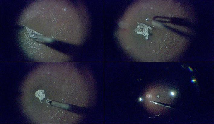
Fig. 2.

Representative intraoperative retinal images of eye with a 441-μm macular hole. Heads-up, 3D system-assisted 27-gauge microincision vitrectomy surgery with minimal illumination was used. The Constellation intraocular illuminator was set to its minimum level, 1%, in all images. Top left: after resecting the vitreal core, we performed triamcinolone acetonide-assisted internal limiting membrane (ILM) peeling. Top right: the ILM was peeled 360 degrees around the macular hole, with its edge attached, and carefully trimmed with a 27-gauge cutter. Bottom left: the ILM flap was inverted and placed over the macular hole. Bottom right: fluid-air exchange was performed with 27-gauge instruments. The macular hole closed completely postoperatively.
