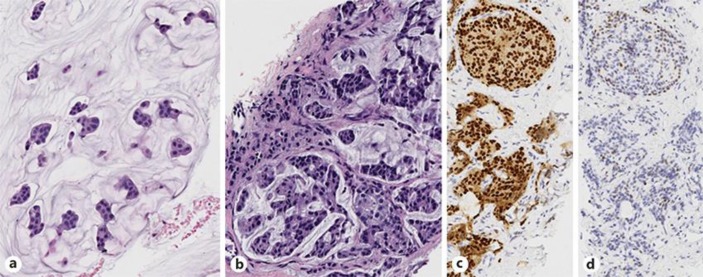
Fig. 1.
a Biopsy of the left breast reveals an extracellular mucin pool containing nests of neoplastic epithelial cells, classic features of mucinous (colloid) carcinoma (HE. ×20). b–d The tumor cells in the CT-guided biopsy of the metastatic liver lesion shows similar morphology to the left breast lesion (b; HE. ×20). A total of 80–90% of tumor cells are strongly positive for ER (c; HE ×20) and ∼5% are positive for PR with low intensity (d; HE ×20). HER2 fluorescence in situ hybridization (FISH) study performed on liver biopsy shows negative HER2/NEU amplification (not shown).

