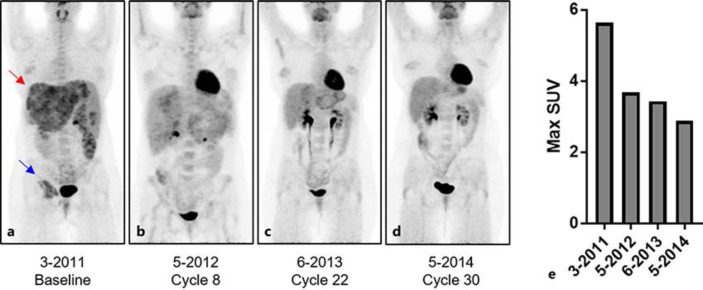
Fig. 2.
a Initial PET scan prior to treatment with doxorubicin shows multiple FDG-avid metastases throughout the liver (red arrow) and a large FDG-avid metastasis in the right acetabulum (blue arrow). b–d PET scans during therapy show decreasing metabolic tumor burden in the liver and in the right acetabulum, achieving a complete metabolic response at both sites by the end of X cycles of doxorubicin. e The maximum standardized uptake value (SUV) of liver metastases decreases from the baseline pretreatment value of 5.6 to an end of treatment maximum SUV of 2.9 which is not significantly different than normal liver SUV.

