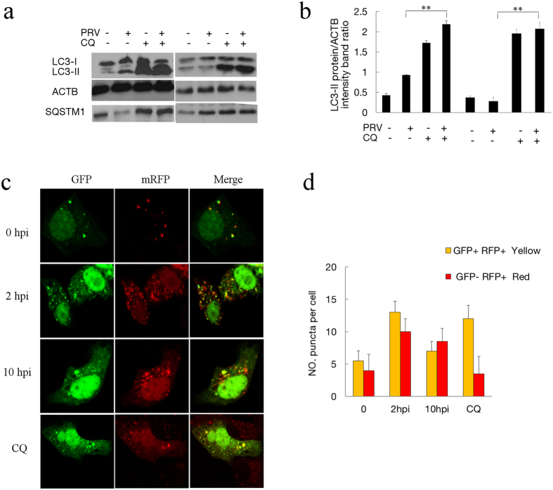
Figure 4. PRV infection enhances autophagy flux.
(a) The modification of LC3 in mock-infected or PRV-infected Vero cells treated with CQ. Vero cells were mock infected or infected with PRV at an MOI of 10. CQ was pretreated for 4 h. At 2 and 8 hpi, cell samples were harvested and lysed, and the cell extracts were analyzed by immunoblotting using anti-LC3 and anti-SQSTM1 antibodies. (b) The ratio of the intensity of LC3-II to ACTB was calculated to represent the autophagic level. The data represent the mean ± SD of three independent experiments. One-way ANOVA test; **P < 0.01. Gels were run under the same experimental conditions. For better clarity and concise presentation, cropped blots are shown. The raw uncropped images can be found in Supplementary Fig. S2. (c) The colocalization analysis of mock-infected Vero cells and cells infected with PRV at an MOI of 10 for different times after transfection with GFP-mRFP-LC3. The cells were fixed and analyzed by confocal microscopy. (d) The graph shows the quantification of autophagosomes and autolysosomes by calculating the average number of dots in 20 cells. Autolysosomes were quantified by counting the number of RFP puncta per cell, and autophagosomes were quantified by counting the number of Yellow puncta per cell. The results are the means of three independent experiments. Error bars indicate the mean ± SD for 20 cells per experimental condition of three independent experiments.

