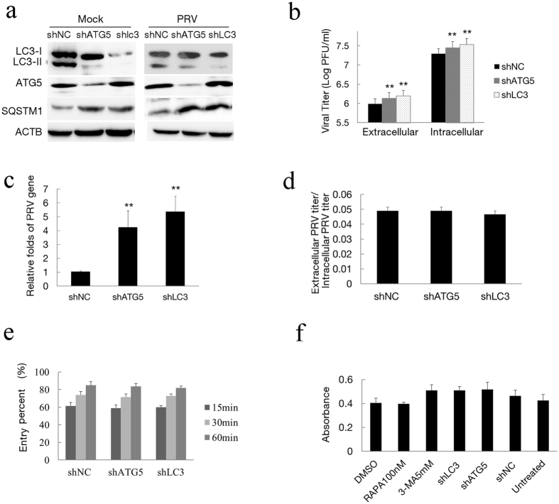
Figure 7. Inhibition of autophagy with specific shRNA targeting endogenous autophagy genes enhances PRV replication.
(a) Vero cells were transfected with shRNAs to knockdown the autophagy proteins ATG5 and LC3B for 24 h. The cells were then mock infected or infected with PRV for 24 h. Western blot analysis was performed to monitor the expression of ATG5 and LC3B protein and autophagic levels. (b) The viral titer determined by PFU assay and the level of the virus gene gB from the treated cells (c) was evaluated by Q-PCR as described in the Methods section. (d) The ratio of the virus titers in supernatants and cell lysates were determined according the results of (b,e) To investigate the effects of ATG5 and LC3B knockdown on virus entry, cells were transfected with shRNA for 24 h and then infected with 100 PFU of PRV. Plaques were counted as described in the Methods section. (f) Cytotoxicity was determined by measuring lactate dehydrogenase (LDH) release from cells with a cytotoxicity assay kit according to the manufacturer’s instructions, and the absorption at 490 nm was measured. Vero cells were transfected with a shRNA vector for 48 h, and a cytotoxicity assay was also performed. One-way ANOVA; *P < 0.05 and **P < 0.01, as compared with the control group. Gels were run under the same experimental conditions. For better clarity and concise presentation, cropped blots are shown. The raw uncropped images can be found in Supplementary Fig. S5.

