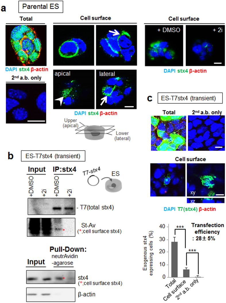
Figure 1. Spontaneous expression of syntaxin-4 at the surface of ES cells.
(a), Endogenous syntaxin-4 (stx4; green) was detected with anti-syntaxin-4 antibodies in ES cells before (Cell surface) and after membrane permeabilization (Total). The cells were precultured in the presence (+2i) or absence (−2i) of GSK3β/MEK1/2 inhibitors for two days. As an internal control, cytoplasmic β-actin was simultaneously detected (red). Left lower column, cells were stained only with secondary antibodies (2nd a.b.). All cells abundantly express β-actin and syntaxin-4 (left). Whereas a small population of the cells cultured in the absence of 2i display cell surface syntaxin-4 at the lateral (arrows) and apical (arrowhead) membrane, cells treated with 2i apparently decrease the extrusion of syntaxin-4 at the cell surface (right images). DMSO alone was used as a control (right images). Cell nuclei were counterstained with DAPI. Bars, 10 μm. (b), ES cells were transfected with syntaxin-4 tagged only with T7 peptide (T7stx4) and surface expression of exogenous syntaxin-4 in cells cultured with (+2i) or without (+DMSO) 2i for two days was analyzed. Upper panel, cell surface proteins were labeled with NHS-sulfo-biotin, and the cell lysate (Input) or immunoprecipitates with anti-syntaxin-4 antibodies (IP:stx4) was analyzed with HRP-labeled anti-T7 antibody (T7) or HRP-labeled streptavidin (St-Av). Lower panel, biotinylated cell surface proteins in the cell lysate (Input) or those retrieved with neutrAvidin-beads (Pull-Down) were analyzed with antibodies against syntaxin-4 or cytoplasmic β-actin. Cropped areas from the original blots (Supplementary Fig. S1) are shown. Treatment with 2i clearly decreases the cell surface expression of T7 tagged-syntaxin-4 (*). (c), ES cells were transfected with T7 tagged syntaxin-4 (T7stx4), and exogenous syntaxin-4 was labeled with anti-T7 tag antibodies (green) after 24 h. As an internal control, cytoplasmic β-actin was simultaneously stained (red). Upper images, anti-T7 tag antibody clearly detects the syntaxin-4 transgene product, but not β-actin, at the cell surface. Bars, 10 μm. Lower panel, the ratio of cells exhibiting syntaxin-4 at the cell surface in transfected cells. Approximately 20% of T7stx4-expressing cells (transfection efficiency, 28 ± 5%) extrudes T7stx4 at the cell surface. N = 18, ***p < 0.001.

