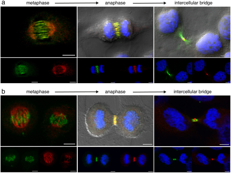
Figure 2. NAIP and the central spindle bundling factors PRC1 and KIF4A.
Confocal differential interference contrast, Hoechst DNA staining, PRC1 or KIF4A (Alexa Fluor-488 shown in green) and NAIP abcam ab25968 (Alexa Fluor-568 shown in red), both individual and merged channels. In the metaphase images the Hoechst channel has been omitted for clarity. (a) PRC1 and NAIP double immunostaining in metaphase showing a predominant presence of NAIP in the spindle poles and PRC1 clearly decorating spindle microtubules. A complete PRC1 and NAIP central spindle colocalization is shown in anaphase. Once the intercellular bridge is formed, PRC1 and NAIP have segregated into the flanking regions and the stem body respectively. (b) KIF4A and NAIP double immunostaining showing a distribution as described in (a) but with a minor presence of KIF4A in the two metaphase spindles shown as well as in the intercellular bridge arms. Bar, 5 μm.

