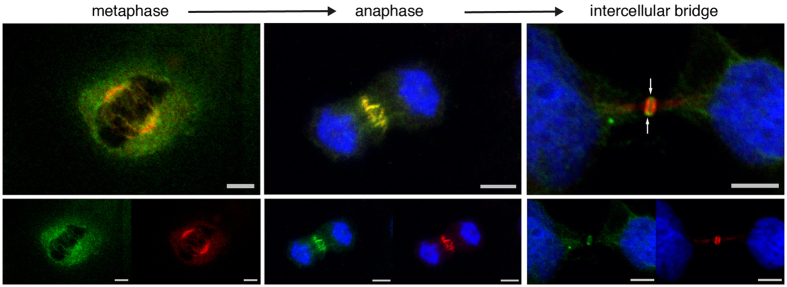
Figure 4. NAIP and Centralspindlin.
Hoechst DNA staining, MgcRacGAP (Alexa Fluor-488 shown in green) and NAIP abcam ab25968 (Alexa Fluor-568 shown in red) confocal channels individually and merged. The Hoechst channel has been omitted in the metaphase images for clarity. In metaphase and anaphase, MgcRacGAP and NAIP largely colocalize (orange in the upper merged images), while in the intercellular bridge MgcRagGAP is predominantly shown in the bulge (arrows) and NAIP is detected in the stem body flanks. Bar, 5 μm.

