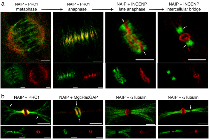
Figure 5. STED microscopy.
Dual STED microscopy for NAIP abcam ab25968 (Chromeo-505 fluorescence shown in red) and PRC1, INCENP or α-Tubulin (Biotin fluorescence shown in green), STED channels merged accordingly. (a) Image series showing the distribution of NAIP at individual phases along the cytokinesis timeline in conjunction with well characterized cytokinesis regulators. Before cytokinesis initiation, in metaphase, NAIP is primarily visualized in the spindle poles, while the microtubule stabilizers (PRC1, KIF4A) and Centralspindlin are observed in spindle microtubules. In anaphase, NAIP immunostaining occupies the center of the central spindle, colocalizes with PRC1, KIF4A, the CPC components and Centralspindlin. Gradual ingression of the cleavage furrow constricts the central spindle into a ring in which NAIP occupies the centermost section (arrows) and colocalizes with MgcRacGAP, while PRC1, KIF4A and CPC have segregated to both sides of NAIP (late anaphase). Then, when the intercellular bridge is completely formed, NAIP is present in the bulge along with MgcRacGAP while the microtubule stabilizers and CPC localize to the intercellular bridge flanking area. (b) NAIP + PRC1: NAIP immunofluorescence is shown in the outer stem body area while PRC1 extends on both sides from the intercellular bridge center well beyond the flanking zone and into the nascent daughter cells (arrows). NAIP + MgcRacGAP: Both proteins are shown delimiting the stem body margins, interestingly MgcRacGAP occupies the upper and lower portions of the stem body precisely (arrows). NAIP + α-Tubulin: An array of microtubule bundles is shown lengthening at both sides on the intercellular bridge center, an abscission point can be identified as a disruption lacking any immunostaining (arrow). Bar, 2, 5 μm.

