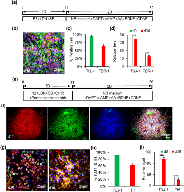
Figure 3. Differentiating iPSCs into cortical and ventral midbrain NSCs in 3D PNIPAAm-PEG hydrogels.
(a) When iPSCs were differentiated in the E6 medium+ LDN193189+ SB431542 in the 3D PNIPAAm-PEG hydrogels for 11 days, and further differentiated in the neural differentiation medium in 2D for 19 days, cortical neurons could be made. 95% and 65% of the produced cells were positive for TUJ-1 and TBR-1, receptively (b,c). (d) qPCR showed the expressions of TUJ-1 and TBR-1 were increased 139 and 51-fold on day 30, respectively. (e) When iPSCs were cultured in the E6 medium + LDN193189 + SB431542 + CHIR99021 + shh + purmorphamine in the 3D PNIPAAm-PEG hydrogels for 11 days, they became FOXA2+ and LMX1A+ ventral midbrain progenitor cells (f). When they were further differentiated in the neural differentiation medium in 2D for 19 days, FOXA2+ and LMX1A+ ventral midbrain neurons could be made. 92% and 63% of the produced cells were positive for TUJ-1 and TBR-1, receptively (g,h). (i) qPCR showed the expressions of TUJ-1 and TH were increased 120 and 25-fold on day 30, respectively. The triple asterisk (***) indicates statistical significance at a level of p < 0.001.

