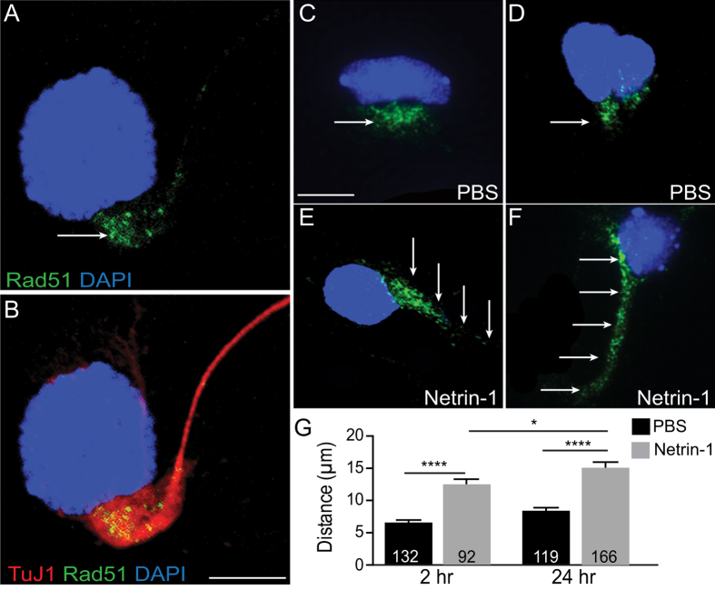
Figure 1. Rad51 was redistributed in cortical neurons in response to Netrin-1.
(A) Representative confocal image of a cultured E14.5 mouse motor cortical neuron immunolabelled for Rad51 (green) and (B) merged with immunolabel for pan neuronal marker TuJ1 (red). Nuclei are counterstained with DAPI (blue). Arrow indicates cytoplasmic Rad51 immunolocalisation aggregated in a peri-nuclear pattern of expression. Scale bar is 5 μm. (C–F) Confocal images of E14.5 mouse motor cortical neurons following treatment with PBS vehicle or Netrin-1 for 2hr (C,E) or 24 hr (D,F). Nuclei are counterstained with DAPI (blue). Arrows depict immunolocalisation of Rad51. Scale bar is 5 μm. (G) Bar graph showing quantification of Rad51 axonal redistribution measured as distance from the nucleus (μm) following 2 or 24 hour treatment with vehicle (PBS) or Netrin-1. Values are mean ± SEM, *p < 0.05, **p < 0.001, ****p < 0.00001.

