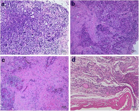
Fig. 2.

Intraoperative findings (microscopy) before the first pressurized intraperitoneal aerosol chemotherapy (PIPAC) (a), and during consecutive PIPAC cycles (PIPAC #3 (b), PIPAC #5 (c), and PIPAC #6 (d). Panel A shows peritoneal manifestations of a serous carcinoma with large, pleomorphic nuclei and frequent mitosis. Histopathological specimens taken during consecutive PIPACs 3, 5, and 6 (panels b, c, and d, respectively) show residual peritoneal tumor foci with reduced cellularity and fibroelastic connective tissue and associated inflammatory changes such as fibrinous exudate, fibrosis, foreign-body reaction, and hemosiderin-laden macrophages. Bars, 100 μm
