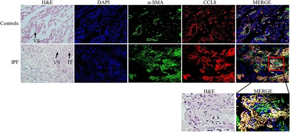
Fig. 4.

Representative double immunofluorescence-stained images of IPF and control lung tissues. CCL8 and α-smooth muscle actin (α-SMA) were stained using PE- (red) and FITC-conjugated antibodies (green), respectively. A proportion of interstitial fibroblasts (IT) and the peribronchial and vascular area (VS) showed staining for both CCL8 and α-SMA (magnification, 200×)
