Fig. 3.

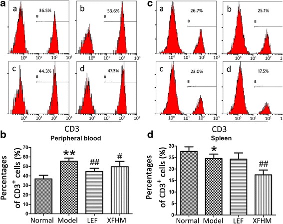
Percentages of CD3+ T cells in peripheral blood cells (PB) and splenocytes. a Histogram of flow cytometry (FCAS): a normal group, b model group, c LEF group and d XFHM group. b The results are presented in the bar charts. Data are presented as the means ± S.D. (n = 6). ** P < 0.01 indicates model group vs. normal group; # P < 0.05 and ## P < 0.01 indicate treatment groups vs. model group
