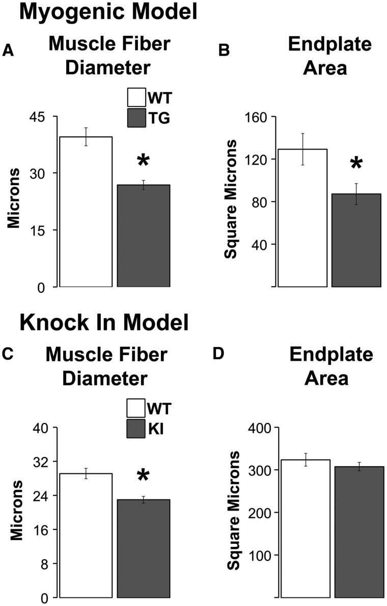
Figure 3.
Endplates are reduced in size only in myogenic TG males but muscle fibres are smaller in diseased males of both models. The size of both fibres and endplates are significantly reduced in TG myogenic males compared to WT males (A, B). While muscle fibres are also smaller in diseased KI males compared to healthy WTs (C), the size of endplates is unaffected by disease in this model (D). This dissociation between endplate and fibre size in diseased KI males raises the possibility that synaptic strength is spared in KI males but direct measures of synaptic strength indicate the opposite, with significant losses in strength in both models (19). * P < 0.05 compared to WT controls. Error bars represent standard error of the mean. n = 4–5 mice/group for estimates of fibre diameter and n = 5–7 mice/group for estimates of endplate area. Twenty junctions were sampled per muscle/animal.

