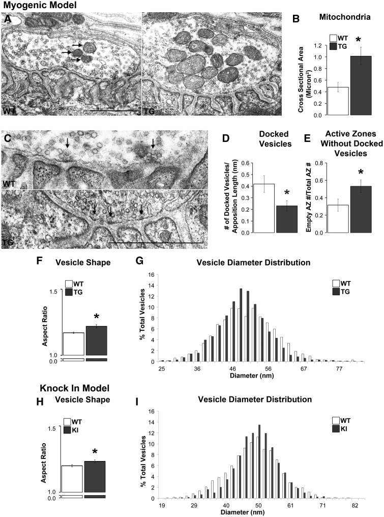
Figure 5.
Disease affects the ultrastructure of motor nerve terminals primarily in myogenic TG males. The cross sectional area of mitochondria (arrows) in TG nerve terminals was significantly larger than in WT terminals (A, B) despite no changes in their number. While the number of active zones, which were identified by an electron dense area directly across from the mouth of a postsynaptic fold (C, denoted with arrows), per length of nerve terminal was unaffected by disease (Table 1), the number of docked vesicles contacting presynaptic membrane per active zone was, with significantly fewer docked vesicles at active zones in TGs compared to WTs (D). Consistent with this finding, the number of active zones without docked vesicles was increased in TG terminals compared to WT terminals (E). Vesicles in TG nerve terminals were also less round than WT vesicles based on their aspect ratio (minimum diameter over maximum diameter; F) although the distribution of average vesicle diameters appeared narrower and more centred around the mean in TG than WT terminals (G). Nerve terminals in KI compared to WT males showed less effects of disease with no difference in the number of active zones, docked vesicles, or active zones without docked vesicles (Table 1). However, vesicles in KI nerve terminals show an increase in their aspect ratio, like TG males, indicating that they are less round than those in WT males (H). Likewise, the distribution of vesicle diameters was narrower and more centred around the mean (I). * P < 0.05 compared to WT controls. Error bars represent standard error of the mean, with n = 30 junctions/group sampled from 3 animals/group, with 10 junctions sampled from each animal. Scale bars represent 1 µm.

