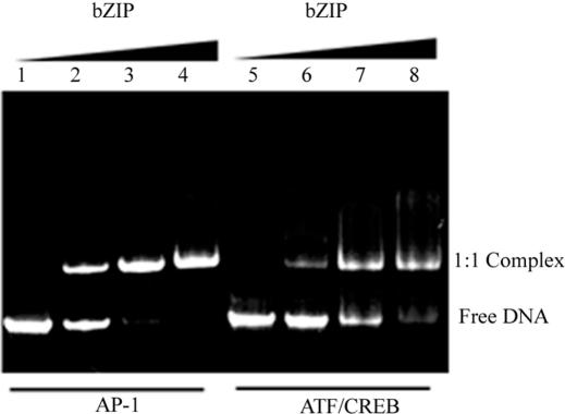
Figure 2.
The PAGE analysis of the constructed U-shaped DNAs and their complexes. Free U-shaped DNA with the AP-1 site (lane 1) and the ATF/CREB site (lane 5) show only one conformation. Lanes 2–4 and lanes 6–8 show binding of the cross-linked bZIP dimer to the DNA having the AP-1 and ATF/CREB sites, respectively. Binding mixtures in 20 μl contained 2.4 μg total DNA. The ratios of bZIP protein to DNA were: 0, 0.6, 1.2 and 1.8. Electrophoresis was on a gradient, 4–20%, non-denaturating polyacrylamide gel at 200 V for 40 min in 20 mM TBE buffer, pH 7.9. For visualization of the bands, the fluorescence of the attached FAM and TAMRA was used.

