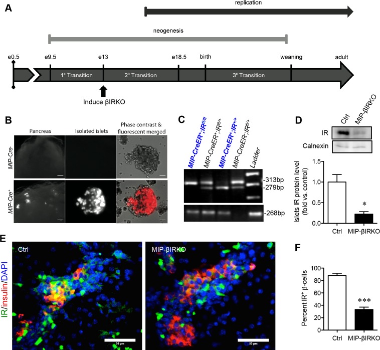
Figure 1. Confirmation of the fetal MIP-βIRKO mouse model.
A. Experimental model schematic with reference timeline of important events during islet development. B. Immunofluorescence image of Cre recombinase expression within pancreatic β-cells of MIP-Cre+, but not MIP-Cre−, mouse islets following cross-breeding to a B6.Cg-Gt(ROSA)26Sortm9(CAG-tdTomato)Hze/J reporter strain. Scale bars: 25 μm (pancreas) and 5 μm (islets). C. Genotypes of fetal mice were determined by PCR of the IR and MIP-CreER genes, followed by subsequent gel electrophoresis. D. Western blotting demonstrated a significant reduction in insulin receptor (IR) protein level of fetal MIP-βIRKO pancreata relative to controls (n = 3-4). IR protein level was normalized to calnexin and expressed as fold vs. controls. Representative blotting is shown. E. Representative double immunofluorescence images and quantification of IR+ β-cells in MIP-βIRKO relative to control pancreatic sections. Scale bar: 50 μm. White bar, control group; black bar, MIP-βIRKO group. Data are expressed as means ± SEM. *p < 0.05, ***p < 0.001 vs. controls. e = embryonic day.

