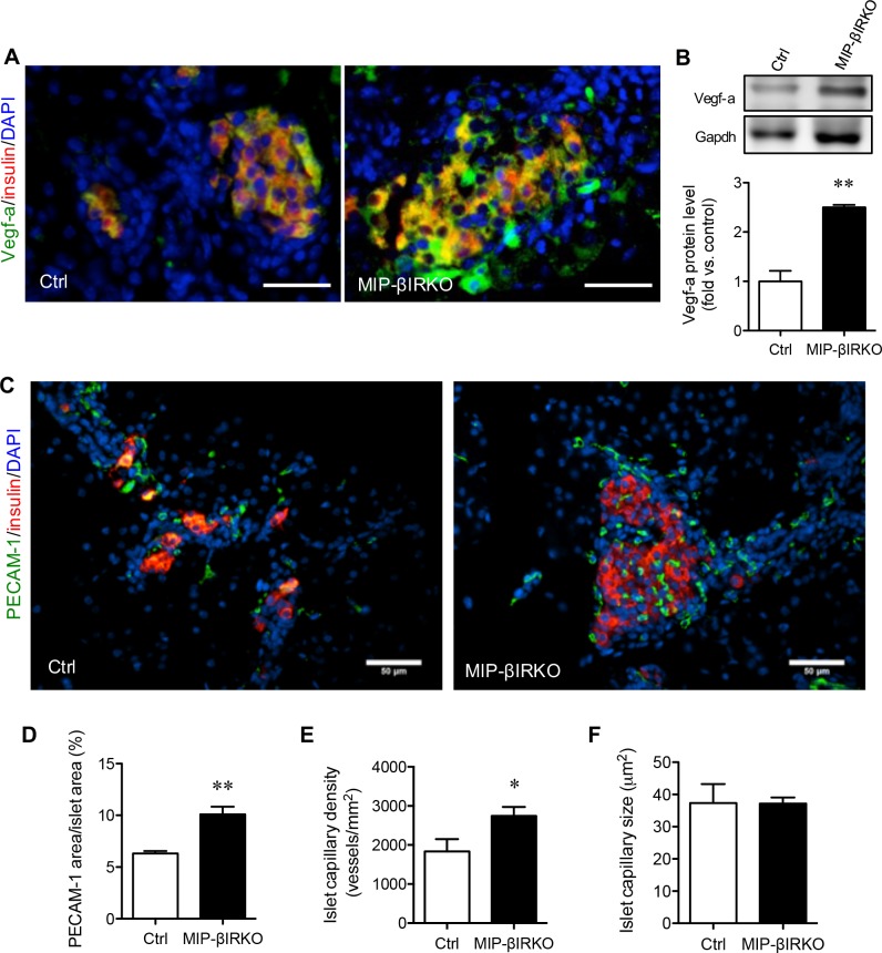
Figure 5. Fetal MIP-βIRKO islets show enhanced islet vascularization.
Representative double immunofluorescence images for A. Vegf-a or C. PECAM-1 (green) with insulin (red), and nuclei stain DAPI (blue), of MIP-βIRKO and control pancreatic sections. Scale bar: 50 μm. B. Western blotting analysis of Vegf-a protein level in fetal pancreas of MIP-βIRKO and control mice (n = 3-5). Representative blotting image is shown. Quantitative analyses of D. percent PECAM-1+ area in the islets, E. islet capillary density and F. average islet capillary size in MIP-βIRKO and controls pancreata. White bars, control group; black bars, MIP-βIRKO group. Data are expressed as means ± SEM (n = 4-5). *p < 0.05, **p < 0.01 vs. controls.

