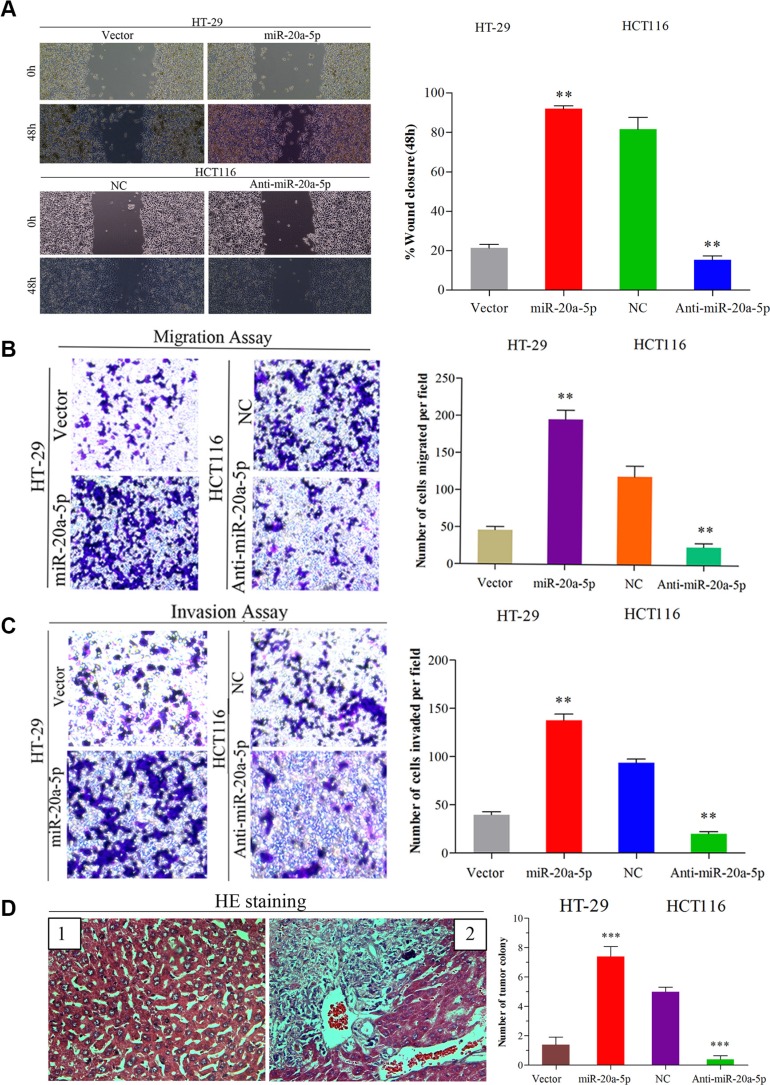
Figure 2. Up-regulated miR-20a-5p promoted the invasion and metastasis of colorectal cancer in vitro and in vivo.
(A) Wound healing assay (B) Migration and (C) invasion assays were performed both in empty vector-infected or miR-20a-5p vector infected HT29 cells and negative control or anti-miR-20a-5p vector infected HCT116 cells, respectively. The results were presented as the mean ± SD of values.(A) The “wound” was created in a cell monolayer, and captured the images at the beginning and at 48 h. Compared the images to quantify the closed rate of the cells,**P < 0.01.(B and C) The number of cells that passed through the membrane was counted in 10 fields (**P < 0.01, magnification ×200). (D) Impact of miR-20a-5p on tumor metastasis in vivo. The number of metastatic liver colonies was counted. The images of normal liver tissue (1) and metastatic colony tissue (2) stained by HE were presented (***P < 0.001, magnification × 200).

