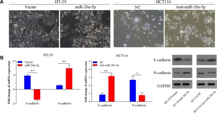
Figure 3. miR-20a-5p promoted the epithelial-to-mesenchymal transition of colorectal cancer cells.
(A) The alteration of phenotypic observed under microscopy (magnification × 200) in miR-20a-5p overexpressing HT29 cells and miR-20a-5p knocking-down HCT116 cells. (B) Expression of E-cadherin and N-cadherin were evaluated at mRNA and protein levels (*P < 0.05, **P < 0.01).

