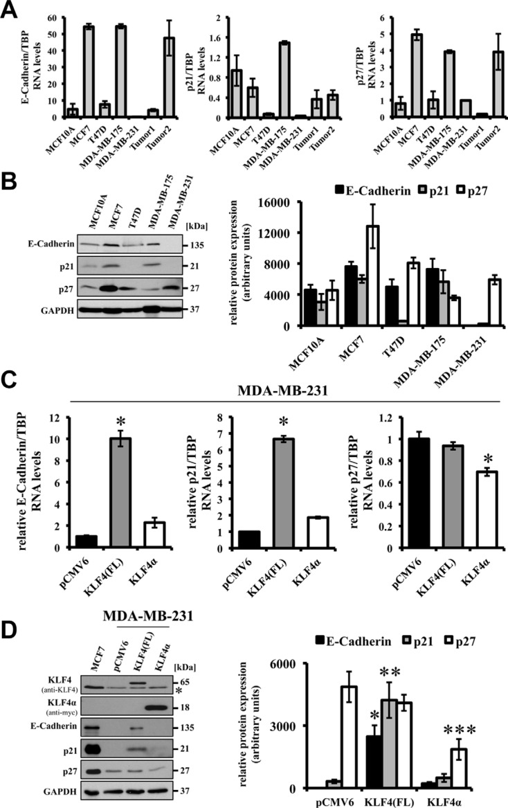
Figure 4. Effects of forced expression of KLF4(FL) and KLF4α in MDA-MB-231 cells.
(A) qPCR analysis of a normal human breast cell line (MCF10A) compared to 4 human breast cancer cell lines and two human ductal breast carcinoma patients for endogenous levels of E-Cadherin, p21Cip1 and, p27Kip1 shows various expression levels in the different samples. Note that E-Cadherin and p21Cip1 levels are very low in MDA-MB-231. Data are expressed as the mean +/− SEM. n = 3. (B) Western Blot analysis of the normal and cancerous breast cells for E-Cadherin, p21Cip1, p27Kip1 indicates that protein levels correlate with RNA levels. Normalized protein levels of three independent western blots have been quantified and plotted. (C) Forced expression of KLF4(FL) in MDA-MB-231 cells induces E-Cadherin as well as p21Cip1 RNA, but not p27Kip1. In contrast, forced expression of KLF4α does neither affect basal levels of E-Cadherin nor p21Cip1, but decreases p27Kip1. Data are expressed as the mean +/− SEM. n = 3. TBP: TATA-Box binding protein. *p ≤ 0.05 (KLF4(FL) versus pCMV6/KLF4α). (D) Response of E-Cadherin, p21Cip1, and p27Kip1 protein levels upon forced expression of KLF4(FL) and KLF4α, respectively, in MDA-MB-231 cells in comparison to the endogenous protein levels in MCF7. Normalized protein levels of three independent western blots have been quantified and plotted. *p ≤ 0.05 E-Cadherin in KLF4(FL) versus KLF4α cells; **p ≤ 0.05 p21Cip1 in KLF4(FL) versus KLF4α cells; ***p ≤ 0.05 p27Kip1 in KLF4α versus KLF4(FL) cells. Note that KLF4α antagonizes KLF4(FL)-mediated target gene regulation and decrease p27Kip1. GAPDH, glyceraldehyde-3-phosphate dehydrogenase. *background band.

